Esophagus Drawing
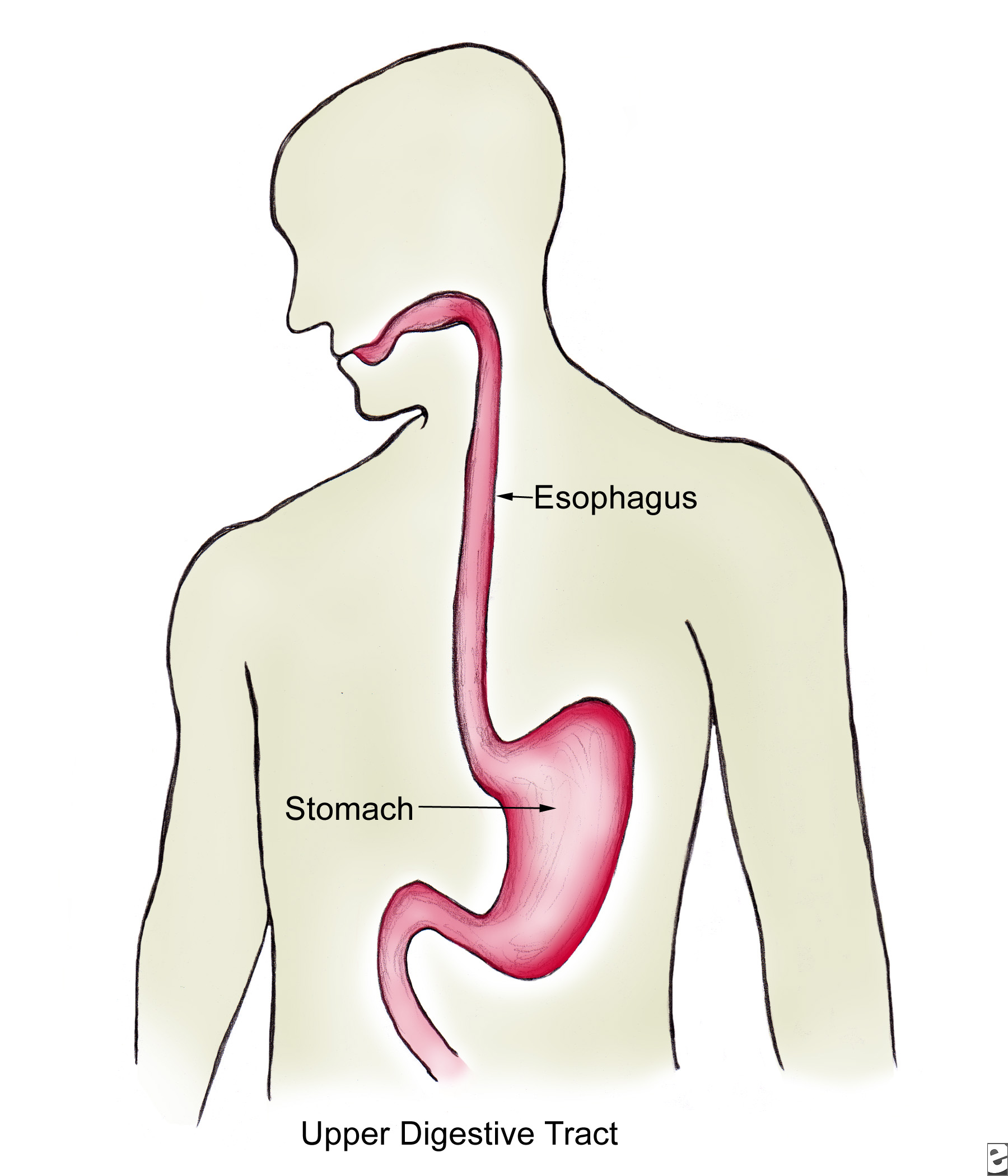
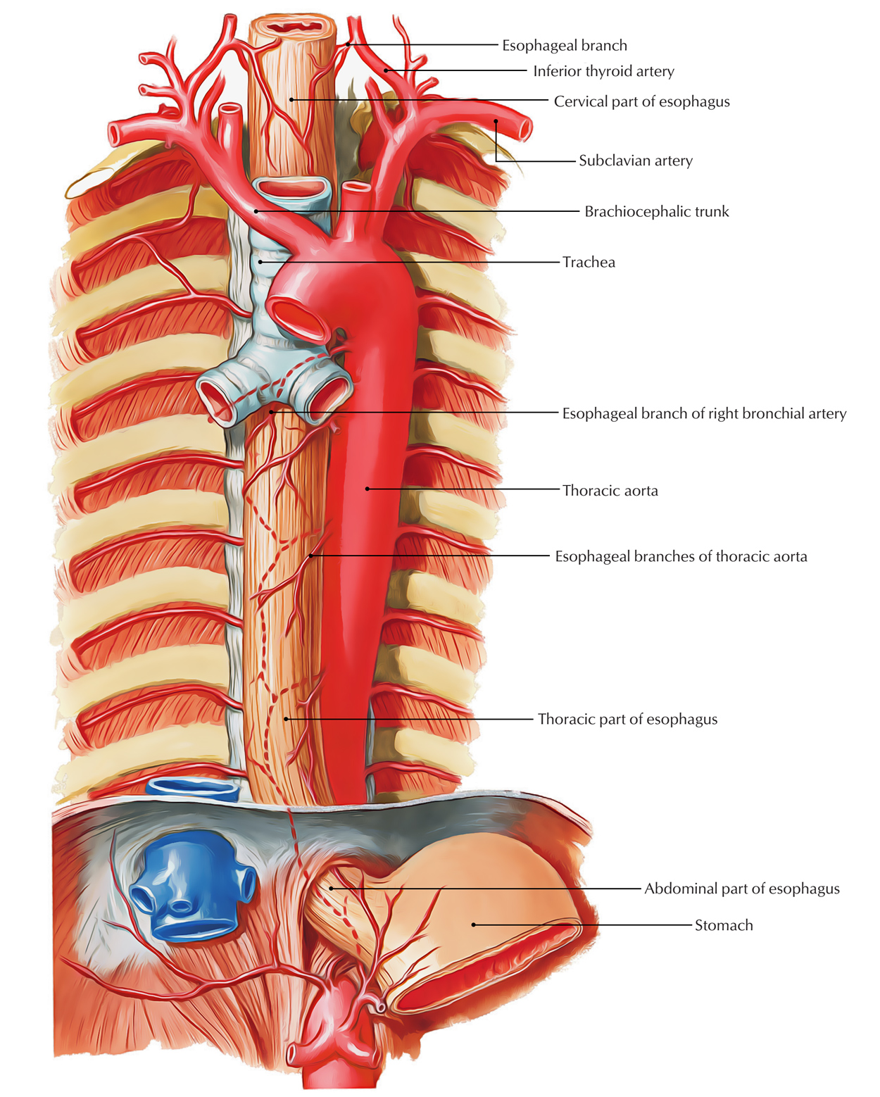
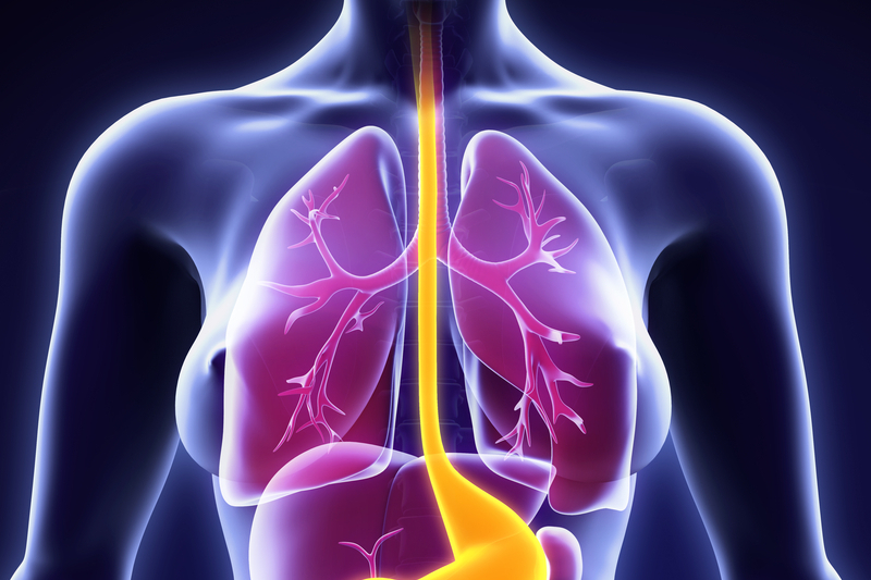
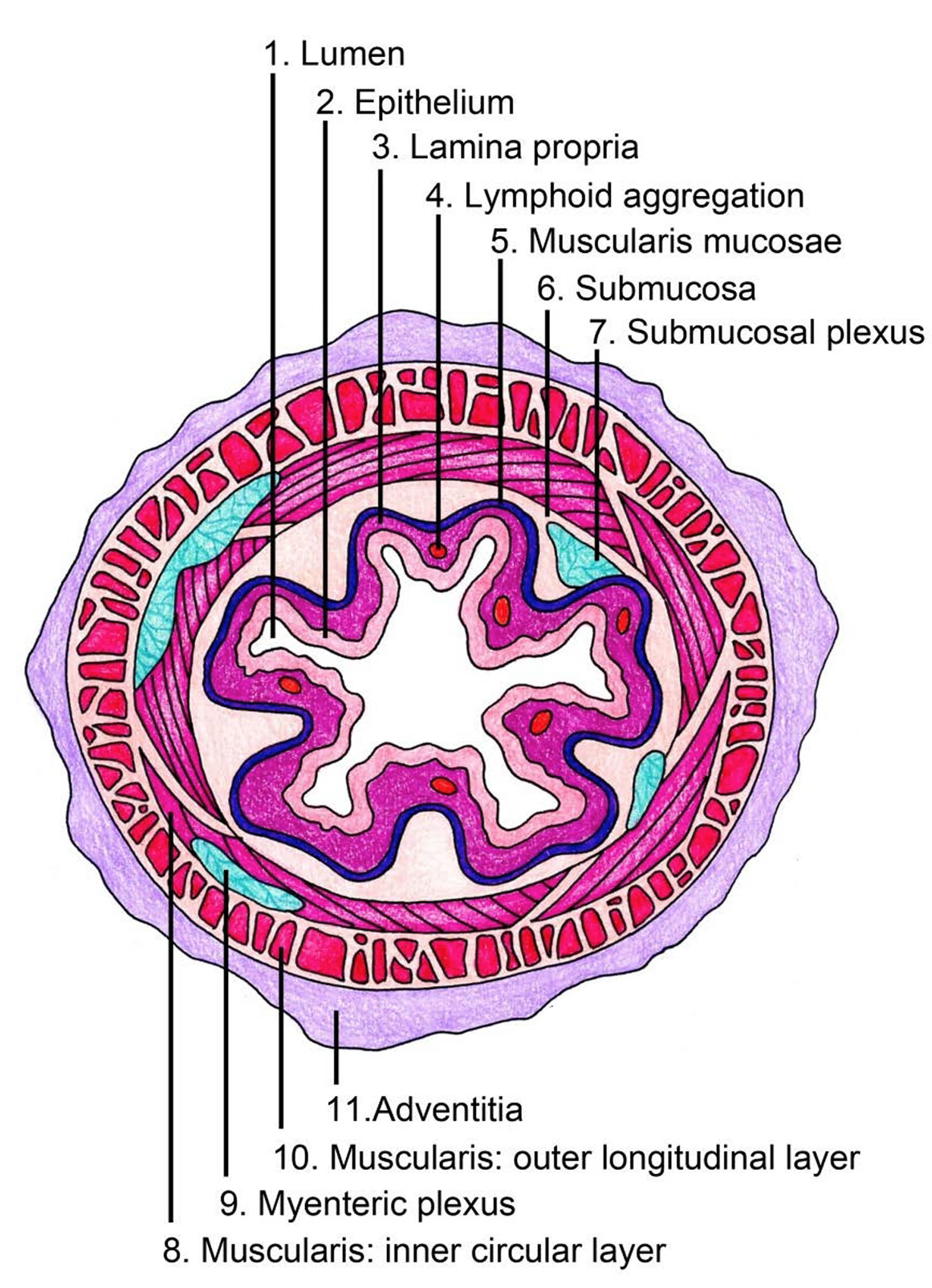
Esophagus Drawing - Do you want to get esophagus histology slide drawing tutorial? A pullout shows the mucosa layer, thin muscle layer, submucosa layer, thick muscle layer, and connective tissue layer of the esophagus wall. Muscles in your esophagus propel food down to your stomach. Most popular smiling stomach color icon smiling stomach color icon. You may follow the same but must try to draw better than this esophagus drawing. Drawing shows the pharynx (throat), esophagus, and stomach. Web anatomy the esophagus is divided into three parts: Web anatomy function associated conditions treatment the esophagus is the muscular tube that connects the back of the throat (or pharynx) with the stomach. Problems with the esophagus include acid reflux and gerd. At the end of the mouth, draw a small tube that extends straight down into the center of your model’s torso. Isolated vector illustration human gut digestive system gastrointestinal tract Problems with the esophagus include acid reflux and gerd. The gastroesophageal junction is noted in this histological section, as shown in the image below. Do you need more images related to esophagus histological. Healthy digestive system, artwork human internal organs icon set Drawing shows the pharynx (throat), esophagus, and stomach. One of the most common symptoms of esophagus problems is heartburn, a burning sensation in the middle of your chest. Most popular smiling stomach color icon smiling stomach color icon. A pullout shows the mucosa layer, thin muscle layer, submucosa layer, thick muscle layer, and connective tissue layer of the esophagus wall.. Its main job is to deliver food, liquids, and saliva to the rest of the digestive system. The lymph nodes are also shown. Tissue around the esophagus is called adventitia. Select from premium esophagus drawing of the highest quality. Cervical which travels through the neck thoracic which is located in the thorax, more specifically in the mediastinum abdominal which travels. Drawing shows the pharynx (throat), esophagus, and stomach. Web browse 1,997 esophagus anatomy photos and images available, or start a new search to explore more photos and images. Healthy stomach inside man body vector illustration. Muscles in your esophagus propel food down to your stomach. You may follow the same but must try to draw better than this esophagus drawing. Web the esophagus is a muscular tube that connects the pharynx to the stomach. Do you need more images related to esophagus histological. Web the esophagus lies posterior to the trachea and the heart and passes through the mediastinum and the hiatus, an opening in the diaphragm, in its descent from the thoracic to the abdominal cavity. Which of the. Web browse 1,997 esophagus anatomy photos and images available, or start a new search to explore more photos and images. Web usmle® step 1 style questions usmle of complete preview a histological section of the esophagus is taken from a healthy patient and examined under microscopy. When we swallow food or liquids the epiglottis falls back and covers the larynx,. Web anatomy the esophagus is divided into three parts: The esophagus has no serosal layer; Most popular smiling stomach color icon smiling stomach color icon. Head section human internal organs. Digestive system male stomach, illustration human anatomy scientific illustrations: Tissue around the esophagus is called adventitia. The lymph nodes are also shown. Isolated vector illustration human gut digestive system gastrointestinal tract Web your esophagus is a hollow, muscular tube that carries food and liquid from your throat to your stomach. Web the esophagus is a hollow tube that begins at the back of the mouth at around the sixth. The gastroesophageal junction is noted in this histological section, as shown in the image below. You may follow the same but must try to draw better than this esophagus drawing. The food moves from the mouth into the esophagus, which carries it down into the stomach. Web the esophagus is a hollow tube that begins at the back of the. Web the esophagus lies posterior to the trachea and the heart and passes through the mediastinum and the hiatus, an opening in the diaphragm, in its descent from the thoracic to the abdominal cavity. Web anatomy function associated conditions treatment the esophagus is the muscular tube that connects the back of the throat (or pharynx) with the stomach. It should. It is approximately 25.4 cm (10 in) in length, located posterior to the trachea, and remains in a collapsed form when not engaged in swallowing. Do you want to get esophagus histology slide drawing tutorial? During swallowing, the epiglottis tilts backwards to prevent food from going down the larynx and lungs. As you can see in figure 23.3.7, the esophagus runs a mainly straight route through the mediastinum of the thorax. A medical anatomy diagram of a woman showing the human digestive system. Web browse 1,997 esophagus anatomy photos and images available, or start a new search to explore more photos and images. Cervical which travels through the neck thoracic which is located in the thorax, more specifically in the mediastinum abdominal which travels past the diaphragm into the abdomen, reaching the stomach cervical part of esophagus pars cervicalis oesophagi 1/3 synonyms: Web esophagitis is an inflammation of the lining of the esophagus, the tube that carries food from the throat to the stomach. Which of the following is true regarding the gastroesophageal junction? Web esophagus histology slide drawing. Web browse 310+ esophagus drawing stock photos and images available, or start a new search to explore more stock photos and images. The esophagus runs behind the trachea. Web anatomy function associated conditions treatment the esophagus is the muscular tube that connects the back of the throat (or pharynx) with the stomach. A pullout shows the mucosa layer, thin muscle layer, submucosa layer, thick muscle layer, and connective tissue layer of the esophagus wall. One of the most common symptoms of esophagus problems is heartburn, a burning sensation in the middle of your chest. Web the esophagus is a hollow tube that begins at the back of the mouth at around the sixth cervical vertebrae.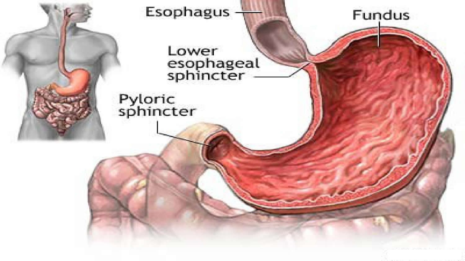
The Human Esophagus Functions and Anatomy and Problems
The esophagus Structure of the esophagus

Esophagus stock illustration. Illustration of throat 40419397
Esophagus Earth's Lab
Esophagus Facts, Functions & Diseases Live Science

esophagus anatomy 2 by speedboy201 on DeviantArt

E.3. Esophagus
The Human Esophagus Functions and Anatomy and Problems

How to Draw Esophagus and Mouth Anotamy Drawing YouTube

The esophagus stock illustration. Illustration of artwork 57504559
Healthy Stomach Inside Man Body Vector Illustration.
Esophagus Drawing Stock Photos Are Available In A Variety Of Sizes And Formats To Fit Your Needs.
Muscles In Your Esophagus Propel Food Down To Your Stomach.
Here In This Section I Am Going To Share Esophagus Slide Image Drawing With You.
Related Post: