Drawing Spinal Cord
Drawing Spinal Cord - While the length of the spinal cord varies from one individual to another, it is usually longer in males (approximately 45 cm) than it is in females (approximately 42 cm). Web each segment of the spinal cord provides several pairs of spinal nerves, which exit from. Spinal cord transevers section diagram to draw easily, it also helpful for reflex arc. It has a relatively simple anatomical course: Uk patients facing weeks of waiting on the nhs are attracted to the private hospital's services. Web the spinal cord is part of the central nervous system (cns), which extends caudally and is protected by the bony structures of the vertebral column. Web the spinal cord begins at the base of the brain and extends into the pelvis. Arterial vasocorona (anastomose between the spinal arteries) They allow you to feel sensations from the external environment (exteroceptive) such as pain, temperature, touch, as well as. The regulation of homeostasis is governed by a specialized region in the brain. Anterior and posterior radicular arteries. It has a relatively simple anatomical course: Also available for free download. Web how to draw a human spinal cord. Describe the type of information carried by each of the three tracts. Black and white vector illustration of children's activity coloring book pages with pictures of orange spine. Eli lew, 35, who works in property development in london,. It then travels inferiorly within the vertebral canal, surrounded by the spinal meninges containing cerebrospinal fluid. Web the spinal cord runs through a hollow case from the skull enclosed within the vertebral column. According. According to some estimates, females have a spinal cord of about 43 centimeters (cm), while males have a spinal cord. Black and white vector illustration of children's activity coloring book pages with pictures of orange spine. Web how to draw a human spinal cord. Draw the location of each of these tracts within the spinal cord. Web 954k views 2. The cerebrum, the diencephalon, the brain stem, and the cerebellum. Brain and spinal cord injuries can also be caused by strokes, infections, and other disorders. Describe the type of information carried by each of the three tracts. Arterial vasocorona (anastomose between the spinal arteries) Web the spinal cord consists of ascending and descending tracts.the ascending tracts are sensory pathways that. Web explore spinal cord diagram with byju’s. Gross anatomy of the spinal cord. The sacral region has a tapered end called the conus medullaris. Web the first section of the cases with drawing version of this module is titled “practice. Three times a week for eight weeks, half of the patients got 20 minutes of the spinal cord stimulation therapy,. Web the olfactory nerve (1st), the optic nerve (2nd), oculomotor nerve (3rd), trochlear nerve (4th), trigeminal nerve (5th), abducens nerve (6th), facial nerve (7th), vestibulocochlear nerve (8th), glossopharyngeal nerve (9th), vagus nerve (10th), accessory nerve (1th), and hypoglossal nerve (12th) 1 comment ( 43 votes) upvote flag Gross anatomy of the spinal cord. Web the spinal cord consists of ascending. Web by the end of this session, learners will be able to: They allow you to feel sensations from the external environment (exteroceptive) such as pain, temperature, touch, as well as. Web © 2023 google llc hi, viewers! Web the vertebral arteries are the main source of blood to the spinal cord. A person’s conscious experiences are based on neural. Three times a week for eight weeks, half of the patients got 20 minutes of the spinal cord stimulation therapy, while the. Spinal cord transevers section diagram to draw easily, it also helpful for reflex arc. Draw the location of each of these tracts within the spinal cord. According to some estimates, females have a spinal cord of about 43. Arterial vasocorona (anastomose between the spinal arteries) Web figure 12.6.1 12.6. Web the spinal cord begins at the base of the brain and extends into the pelvis. Black and white vector illustration of children's activity coloring book pages with pictures of orange spine. Web 954k views 2 years ago. The spinal cord is divided into four regions: Web the vertebral arteries are the main source of blood to the spinal cord. Web the spinal cord begins at the base of the brain and extends into the pelvis. Describe the type of information carried by each of the three tracts. Also available for free download. Web the spinal cord begins at the base of the brain and extends into the pelvis. The bundle of axons inferior to the conus medullaris is the cauda equina. Brain and spinal cord injuries can also be caused by strokes, infections, and other disorders. Web how to draw a simple diagram of human spinal cord uc easy drawings 8.35k subscribers subscribe subscribed 3 share 340 views 1 year ago #howtodraw #drawings #draw how to draw human. Eli lew, 35, who works in property development in london,. How to draw human backbone (spine) drawing with sketch pen and pencilsubscribe to my channel to ge. Web the spinal cord is a single structure, whereas the adult brain is described in terms of four major regions: Web the spinal cord runs through a hollow case from the skull enclosed within the vertebral column. Many of the nerves of the peripheral nervous system, or pns, branch out from the spinal cord and travel to. Web the spinal cord is a cylindrical mass of neural tissue extending from the caudal aspect of the medulla oblongata of the brainstem to the level of the first lumbar vertebra (l1). Here i upload carefully crafted videos to meet the problems in drawing.all the videos are. Anterior and posterior radicular arteries. Web the spinal cord consists of ascending and descending tracts.the ascending tracts are sensory pathways that travel through the white matter of the spinal cord, carrying somatosensory information up to the brain. Web the olfactory nerve (1st), the optic nerve (2nd), oculomotor nerve (3rd), trochlear nerve (4th), trigeminal nerve (5th), abducens nerve (6th), facial nerve (7th), vestibulocochlear nerve (8th), glossopharyngeal nerve (9th), vagus nerve (10th), accessory nerve (1th), and hypoglossal nerve (12th) 1 comment ( 43 votes) upvote flag Web each segment of the spinal cord provides several pairs of spinal nerves, which exit from. Welcome to my just made easy official youtube channel.
Spinal Cord Diagram with Detailed Illustrations and Clear Labels

Spinal Cord Anatomy Nurse Info
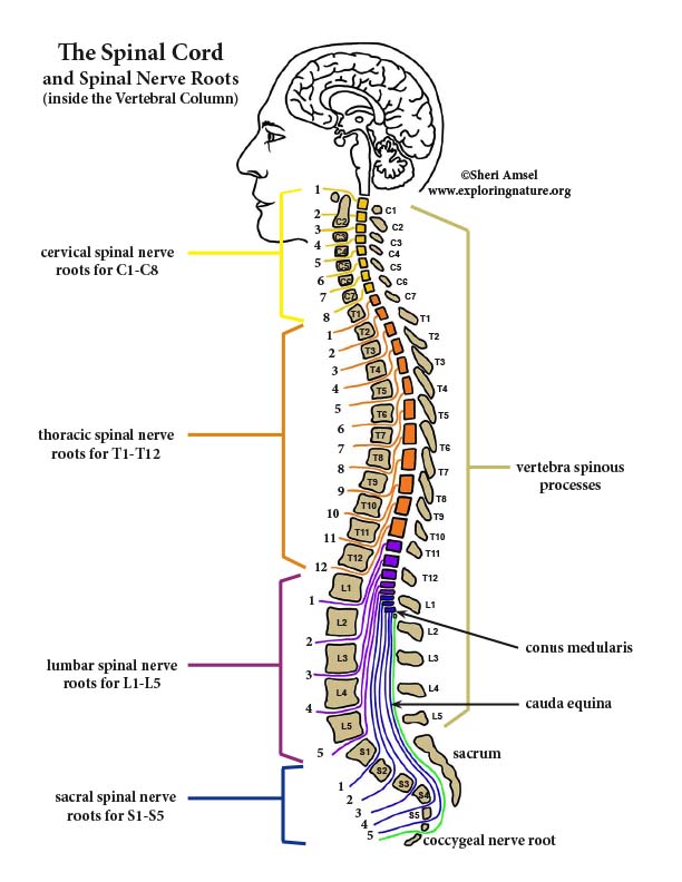
Spinal Cord and Spinal Nerve Roots Diagram
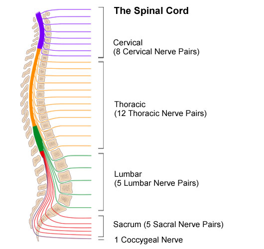
Anatomy of the Spinal Cord

The Spinal Cord Neurologic Clinics
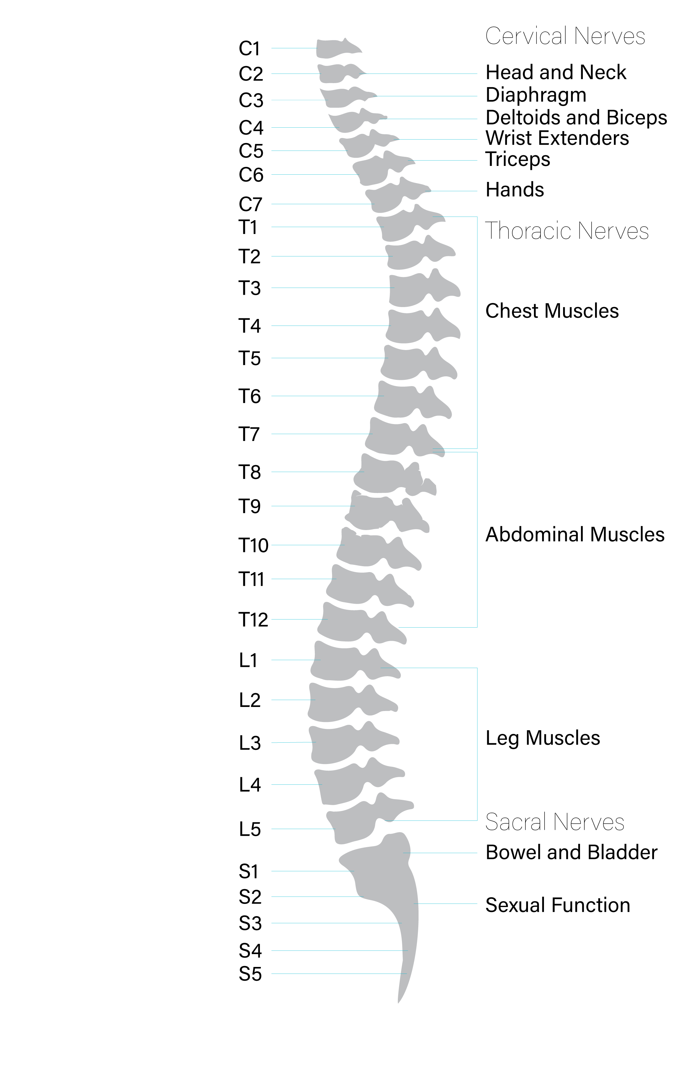
Anatomy of the Spinal Cord Praxis Spinal Cord Institute

How to Draw Structure Of The Spinal Cord Diagram Easy And Step by Step
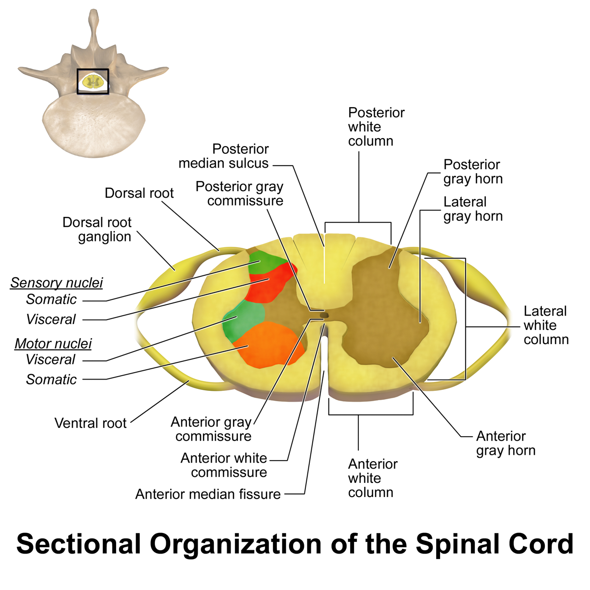
Spinal Cord Summary Neuroanatomy Geeky Medics
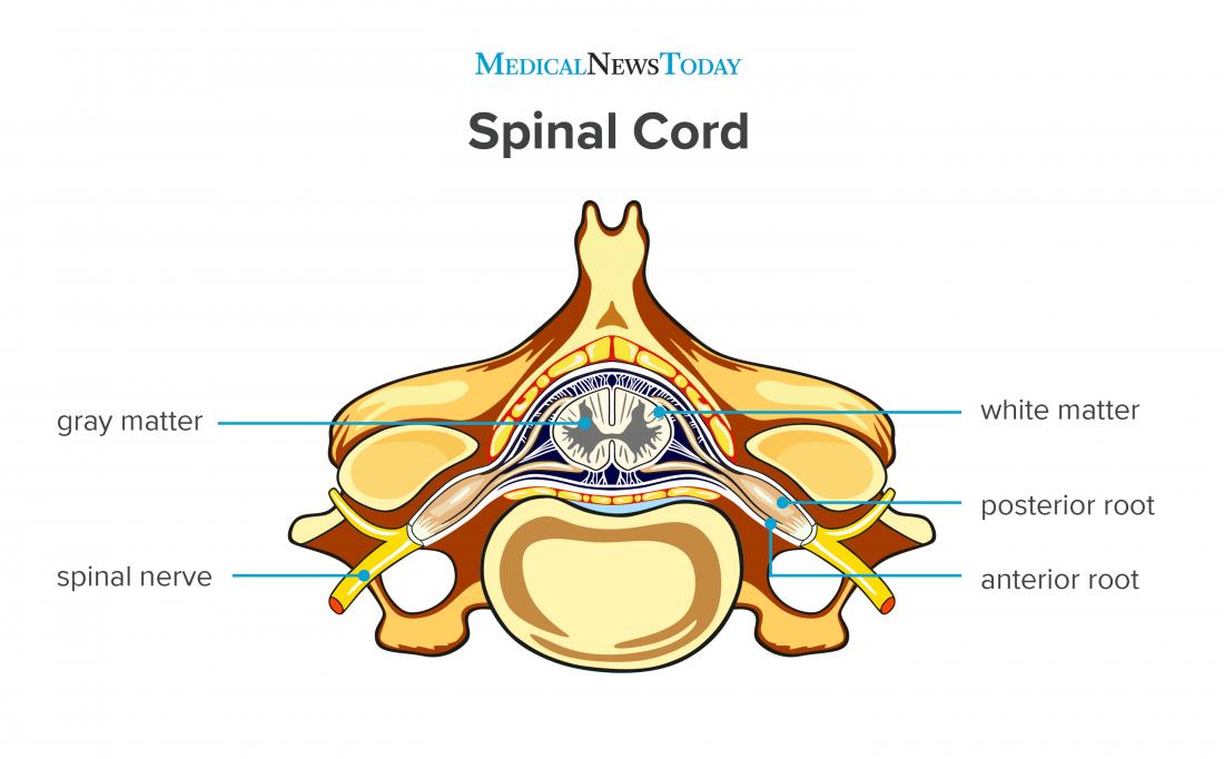
Spinal cord Anatomy, functions, and injuries

Spinal Cord Labeled
Web How To Draw A Human Spinal Cord.
They Allow You To Feel Sensations From The External Environment (Exteroceptive) Such As Pain, Temperature, Touch, As Well As.
Web © 2023 Google Llc Hi, Viewers!
The Spinal Cord Is Divided Into Four Regions:
Related Post: